See Explanation
/uploads/2678_body_ebb50ee6fedf0796bd2fa51a14ff6c95.jpg
Ophthalmology
infectious conditions
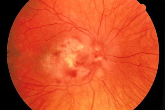
cytomegalovirus retinitis
A 44-year-old man with a documented history of human immunodeficiency virus (HIV) infection presents to an outpatient clinic with a two-week history of progressively worsening blurred vision in his right eye, accompanied by new onset of "cobwebs" and "spider-like" floaters. He admits to a significant lapse in his antiretroviral therapy for over five years and has not had a follow-up with an infectious disease specialist during this period. On ophthalmic examination, visual acuity in the right eye is 20/200, and the left eye is 20/25. Slit-lamp examination of the anterior segment is unremarkable. Fundoscopic examination of the right eye reveals extensive areas of peripheral retinal whitening with associated hemorrhage, consistent with a 'cottage cheese and ketchup' appearance, along the vascular arcades. The left fundus appears normal. Given these findings, what is the most likely diagnosis and the crucial initial management approach?
| Lab Parameter | Value | Reference Range |
|---|---|---|
| White Blood Cell Count | 4.5 x 10^9/L | 4.0-11.0 x 10^9/L |
| Hemoglobin | 138 g/L | 130-170 g/L |
| CD4+ T-cell count | 38 cells/µL | 500-1500 cells/µL |
| HIV-1 RNA (Viral Load) | 185,000 copies/mL | Undetectable to < 50 copies/mL |
Edit question